A diagnosis of mesothelioma is most often obtained with careful assessment of clinical and radiological findings in addition to a confirming tissue biopsy. (Learn about typical mesothelioma symptoms.) A review of the patient's medical history, including history of asbestos exposure is taken, followed by a complete physical examination, x-rays of the chest or abdomen, and lung function tests. A CT scan or MRI may also be done at this time. If any of these preliminary tests prove suspicious for mesothelioma; a biopsy is necessary to confirm this diagnosis.
Imaging Techniques and Their Value in Diagnosing and Assessing Mesothelioma.
There are several imaging techniques which may prove useful when mesothelioma is suspected due to the presence of pleural effusion combined with a history of occupational or secondary asbestos exposure. While these imaging techniques can be valuable in assessing the possibility of the cancer, definitive diagnosis is still most often established through fluid diagnosis or tissue biopsy.
Some of the most commonly used imaging methods include:
• X-ray
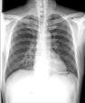
A chest x-ray can reveal pleural effusion (fluid build-up) which is confined to either the right (60%) or left (40%) lung. On occasion, a mass may be seen. Signs of prior non-cancerous asbestos disease, such as pleural plaques or pleural calcification, or scarring due to asbestosis may also be noted.
• Computed Tomography (CT)
CT scans are also able to define pleural effusion, as well as pleural thickening, pleural calcification, thickening of interlobular fissures, or possible chest wall invasion. CT, however, is not able to differentiate between changes associated with benign asbestos disease (pleural disease), or differentiate between adenocarcinoma of the lung wh
ich may have spread to the pleura verses mesothelioma. CT scans may also be valuable in guiding fine needle aspiration of pleural masses for tissue diagnosis.
• Magnetic Resonance Imaging (MRI)
MRI scans are most often used to determine the extent of tumor prior to aggressive treatment. Because they provide images in multiple planes, they are better able to identify tumors as opposed to normal structures. They are also more accurate than CT scans in assessing enlargement of the mediastinal lymph nodes (those lymph nodes which lie between the two lungs), as well as a clear diaphragmatic surface, both of which play an important role in surgical candidacy.
• Positron Emission Tomography (PET)
PET imaging is now becoming an important part of the diagnosis and evaluation of mesothelioma. While PET scans are more expensive than other types of imaging, and are not always covered under insurance, they are now considered to be the most diagnostic of tumor sites, as well as the most superior in determining the staging of mesothelioma. Further explanation of PET scans.
• CT/PET
For patients who may be candidates for aggressive multimodality treatment (surgery, chemotherapy and radiation), accurate clinical staging is extremely important. Integrated CT/PET imaging provides a relatively new tool in this respect, and has become the imaging technique of choice for determining surgical eligibility. By combining the benefits of CT and PET (anatomic and metabolic information) into a single scan, this technology can more accurately determine the stage of the cancer, and can help identify the best treatment option for the patient. Read about a study of CT-PET imaging in preoperative evaluation of patients with malignant pleural mesothelioma.
A needle biopsy of the mass, or the removal and examination of the fluid surrounding the lung, may be used for diagnosis, however, because these samples are sometimes inadequate as far as determining cell type (epithelial, sarcomatous, or mixed) or because of the unreliability of fluid diagnosis, open pleural biopsy may be recommended. In a pleural biopsy procedure, a surgeon will make a small incision through the chest wall and insert a thin, lighted tube called a thoracoscope into the chest between two ribs. He will then remove a sample of tissue to be reviewed under a microscope by a pathologist. In a peritoneal biopsy, the doctor makes a small incision in the abdomen and inserts a peritoneoscope into the abdominal cavity.
Once mesothelioma is suspected through imaging tests, it is confirmed by pathological examination. Tissue is removed, put under the microscope, and a pathologist makes a definitive diagnosis, and issues a pathology report. This is the end of a process that usually begins with symptoms that send most people to the doctor: a fluid build-up or pleural effusions, shortness of breath, pain in the chest, or pain or swelling in the abdomen. The doctor may order an x-ray or CT scan of the chest or abdomen. If further examination is warranted, the following tests may be done:
- Video-Assisted Thoracoscopic Surgery (VATS)
Over the past decade, the use of video-assisted thoracic surgery (VATS) has become one of the most widely used tools in the diagnosis of mesothelioma. Biopsies of the pleural lining, nodules, masses and pleural fluid can now easily be obtained using this minimally invasive procedure, and other therapies such as pleurodesis (talc) for pleural effusions can be done concurrently.While the patient is under general anesthesia, several small incisions or “ports” are made through the chest wall. The surgeon then inserts a small camera, via a scope, into one incision, and other surgical instruments used to retrieve tissue samples into the other incisions. By looking at a video screen showing the camera images, the surgeon is able to complete whatever procedures are necessary
In many cases, this video-assisted technique is able to replace thoracotomy, which requires a much larger incision to gain access to the chest cavity, and because it is minimally invasive, the patient most often has less post-operative pain and a potentially shorter recovery period.
For pleural mesothelioma the doctor may look inside the chest cavity with a special instrument called a thoracoscope. A cut will be made through the chest wall and the thoracoscope will be put into the chest between two ribs. This test is usually done in a hospital with a local anesthetic or painkiller.
If fluid has collected in your chest, your doctor may drain the fluid out of your body by putting a needle into your chest and use gentle suction to remove the fluid. This is called thoracentesis.
For peritoneal mesothelioma the doctor may also look inside the abdomen with a special tool called a peritoneoscope. The peritoneoscope is put into an opening made in the abdomen. This test is usually done in the hospital under a local anesthetic.
If fluid has collected in your abdomen, your doctor may drain the fluid out of your body by putting a needle into your abdomen and using gentle suction to remove the fluid. This process is called paracentesis.
If abnormal tissue is found, the doctor will need to cut out a small piece and have it looked at under a microscope. This is usually done during the thoracoscopy or peritoneoscopy, but can be done during surgery. More on needle biopsies.
Pathology and The Role of Pathologists in the Diagnostic Process.
Pathology, or the scientific study of cells, tissue, or fluid taken from the body is an integral part of a mesothelioma diagnosis. Most hospitals have their own pathology labs staffed by board-certified pathologists and licensed technologists. The importance of pathological diagnosis can not be underestimated, since the course of treatment is dependent upon an accurate diagnosis.
To make a diagnosis, pathologists examine tissue under a microscope, and based on established criteria, make a determination of benign vs. malignant cells. (More on biopsy tissue processing.) Subsequently, the type of cancer is determined. Although most pathologists have a general expertise of various diseases, a small number acquire training in a subspecialty, such as mesothelioma. These are physicians who have received world-wide recognition as premier experts, and have achieved high acclaim for their research, published articles and abstracts, and teaching. For a list of expert pathologists in the field of mesothelioma diagnosis, please call the MW toll free at 1-877-367-6376 or fill in the form at the bottom of this page specifying your request.
Knowing the stage is a factor in helping the doctor form a treatment plan. Mesothelioma is considered localized if the cancer is confined to the pleura, or advanced if it has spread beyond the pleura to other parts of the body such as the lungs, chest wall, abdominal cavity, or lymph nodes.
Immunohistochemical Markers for Mesothelioma.
A diagnosis of any specific type of cancer often means ruling out other cancers in the process. This is true in the case of mesothelioma, where the most common “differential diagnosis” is that of adenocarcinoma versus mesothelioma.
During the biopsy procedure, the surgeon removes tissue samples to be sent to the laboratory. In the lab, slides are produced and then viewed and analyzed by a pathologist. These tissue specimens arrive at the lab with a request form that details patient information and history along with a description of the site in the body from which the specimen was obtained. Each individual specimen is numbered for each patient.
The pathologist then does a “gross examination” which consists of describing the tissue, and then placing it in a plastic cassette. The cassettes are then placed in a fixative that preserves the tissue permanently. Once the tissue has been fixed, it is processed into a paraffin block that will allow the pathologist to slice off thin microscopic sections that will then be stained to determine the patient’s diagnosis.
Immunohistochemistry is defined as “a method of analyzing and identifying cell types based on the binding of antibodies to specific components of the cell”. It is this process that helps diagnose mesothelioma versus adenocarcinoma (or other types of cancer).
Early on, the “markers” which helped distinguish mesothelioma from adenocarcinoma were “negative markers”; those expressed in adenocarcinomas, but not in mesotheliomas. This made it more difficult to confirm a diagnosis, because pathologists were dealing with the absence of, rather than the presence of certain markers. Some of these markers, which are normally “positive” in an adenocarcinoma diagnosis and “negative” in a mesothelioma diagnosis, are carcinoembryonic antigen (CEA), CD 15 (LeuM1), epithelial glycoprotein (Bg8), tumor glycoprotein (BerEp4) and tumor glycoprotein (MOC-31).
In more recent years, “positive markers” expressed by mesotheliomas have come to the forefront. Some of the markers which are normally “positive” in mesotheliomas and “negative” in adenocarcincomas are calretinin, cytokeratin 5, HBME-1, mesothelin, N-cadherin, thrombomodulin, vimentin and Wilm’s tumor gene product (WT-1).
It is important to remember that while the above markers are commonly used to help diagnose the epithelial sub-type of mesothelioma, that they may also be expressed in other types of cancer, and may not necessarily apply to the bi-phasic or sarcomatoid sub-types of mesothelioma. Your doctor can always contact a more specialized lab if he/she feels your diagnosis is in any way inconclusive.
FOR YOUR FURTHER READING.............
Mesothelioma Diagnosis and Mesothelioma Detection
» Mesothelioma Imaging Technologies
» Diagnostic Biopsy Surgery
» New Methods for Detection
» New Blood Tests
Malignant mesothelioma detection, like other cancers, can be accomplished with imaging equipment, such as x-ray machines. But once detected, mesothelioma diagnosis is difficult for a number of reasons. First, there is a very extended time period between the exposure to asbestos and the onset of the disease, sometimes as long as 50 to 60 years. Patients would often not think to tell their doctors about working in an asbestos-related job many years earlier.
Second, the typical symptoms of mesothelioma, shortness of breath and coughing, are also symptoms of many other types of lung problems, both cancerous and non-cancerous. Thus, just because a person has these symptoms, it does not in any way provide a mesothelioma diagnosis.
Third, many types of tumors can exist in the serous cavities that are not mesothelioma. These other types of tumors can be non cancerous, or benign, that originate in the tissues of the serous membranes, other than the mesothelium. Or they can be tumors that have migrated from other organs with cancerous growths due to metastases.
Mesothelioma and Imaging Technologies.
X-rays and other types of imaging technologies can be used to detect tumors or effusion (build up of fluid) in the body, including mesothelioma detection. A growth in the chest cavity will show up in an X-ray or MRI analysis. But these devices cannot directly determine the type of cancer or provide a mesothelioma diagnosis. They cannot determine whether the tumor is mesothelioma or originates from some other source.
Positron emission tomography (PET) is a type of diagnostic imaging scan that is used for malignant mesothelioma detection. PET scans use the emission of positrons (tiny particles that are emitted from radioactive substances) for the purpose of radiation detection.
Some medical professionals are of the impression that PET scans are the most effective method through which to definitively verify a case of mesothelioma. While they believe that standard imaging techniques like x-rays and MRIs should continue to play a role in diagnosing the disease, it is felt that positron emission tomography is becoming an increasingly valuable tool in the staging and typing of the latent asbestos cancer.
Diagnostic Surgery - Biopsy.
To provide a mesothelioma diagnosis, a biopsy is needed. This biopsy then undergoes what is called diagnostic histopathology. Histopathology is a technique where the cells from the tumor are viewed under a high-powered microscope, or electron microscopy. Electron microscopy is considered the gold standard for evaluating tumor material from a biopsy. It is a highly advanced microscope that allows viewing of the tiniest elements of cell tissue.
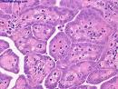
For mesothelioma diagnosis, a pathologist (a doctor who specializes in disease detection) places the tumor cells in the electron microscope and then views the structure of the individual cells. The mesothelioma cells have a specific shape and pattern, and this allows them to be identified by a very keen pathologist. But mesothelioma cells also look similar to other types of cancer cells, such as adenocarcinoma cells, and this can make the pathologist's job very difficult. Even with the electron microscope, the different types of mesothelioma cells can be hard to recognize. The three types of cells are epithelioid mesothelioma cancer cells, which are tubular in shape, sarcomatoid mesothelioma cancer cells, which are oval and irregularly shaped, and biphasic mesothelioma cancer cells, which are a combination of shapes. These cells can be confused with other types of cancer cells.
New Methods For Mesothelioma Detection.
Due to this diagnostic confusion, much research is underway to find new methods for diagnosis. One method is to evaluate the types of compounds generated by the mesothelioma cancer cells. This is called histochemistry. Histochemical reactions have long been used to distinguish between mesothelial and other types of tumor cells. For example, mesothelial cells are known to produce specific types of carbohydrate compounds. Unfortunately, other types of cells in the body also produce these compounds.
Immunochemistry is also being used to detect mesothelioma. This area of study evaluates the presence of antibodies in the body. Certain types of antibodies are known to be associated with certain types of cancer. But mesothelial cells have no specific types of antibodies that can provide a "positive" marker. Consequently, immunochemistry allows the doctor to "eliminate" the other cancers, but does not indicate the presence of mesothelioma. These techniques offer insight into the disease and may help eliminate other diseases, but none can directly detect mesothelioma.
New Blood Tests for Mesothelioma Detection.
SMR Protein: Recently, because of the difficulty in diagnosing malignant mesothelioma, research has concentrated on finding new ways to detect the presence of the disease. Researchers in Australia have found that a certain protein, called SMR or Soluble Mesothelin Related protein, is elevated in patients with mesothelioma. These researchers have suggested that a test for the presence of SMR in the blood could represent a useful marker for the diagnosis and disease progression. They feel that such a diagnosis tool could lead to earlier detection, and thus more effective treatment.
One of the most striking findings of their research was that several asbestos-exposed persons who tested positive for SMR were diagnosed with mesothelioma within three years. They suggested that evaluation of SMR may help to identify persons at risk for this deadly disease. Also, they found that SMR levels increase as mesothelioma progresses, suggesting that SMR evaluation could be used to track the progression of the disease and the effectiveness of treatment
Osteopontin Glycoprotein: In an effort to produce the first early-detection test to screen for malignant mesothelioma, researchers at Wayne State University have been studying the possible link between mesothelioma development and levels of a glycoprotein called osteopontin. Early clinical study findings of 190 patients have demonstrated a link between high levels of osteopontin and the development of malignant pleural mesothelioma.
Although the results are being viewed as preliminary, there is a great deal of excitement surrounding the potential of a blood test capable of screening for mesothelioma in its earliest stages. While there is no known cure for malignant mesothelioma, research is ongoing and certain successes have already been realized in terms of extending survival time beyond the one to two year post-diagnosis average. It is hoped that if mesothelioma specialists have more time through which to conduct treatment on a lesser developed form of the asbestos cancer, the greater a patient's chance at potential survival.
Despite the fact that the preliminary results of the osteopontin blood test clinical trial have been met with some controversy, the National Cancer Institute (NCI) continues to sponsor additional study.
Tell Your Doctor About Asbestos Exposure.
If you or a loved one has been exposed to asbestos, even if it was in the distant past, it is very important that you inform your doctor. One reason why mesothelioma is such a deadly disease is that it is detected late in the disease process. If your doctor knows of the exposure, he or she may be more aware of your symptoms or other health issues that could be used for early detection.